
Fig. 2.
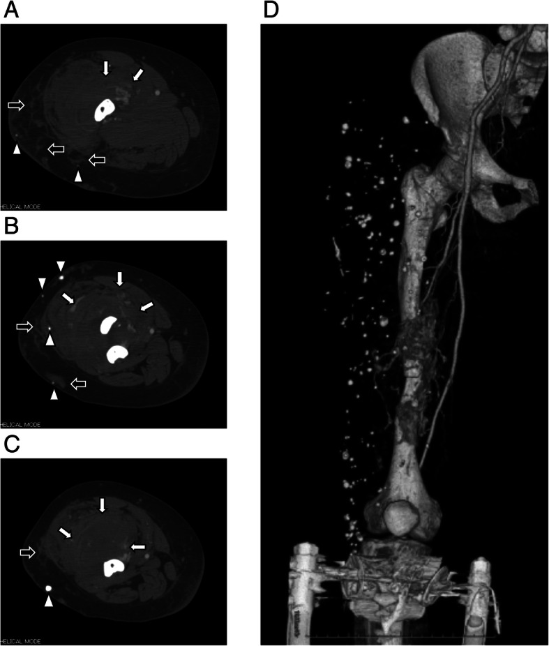
Preoperative enhanced computed tomography scans show large arteriovenous malformations (AVMs) in the quadriceps femoris (white arrows), which are partially present subcutaneously (black arrows). Numerous phleboliths (white arrowheads), massive bony erosion, and very narrow intramedullary canal are also observed (a: proximal site of fracture; b: fracture site; c: distal site of fracture). d Three-dimensional computed tomography angiography scan shows the AVMs are fed by the branches of the deep and superficial femoral artery