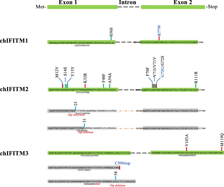
Fig. 4.
SNVs and INDELS localization across the chIFITM proteins. chIFITM proteins are comprised of two exons separated by one intron. SNVs and INDELS locations from Table 3 are depicted in red (missense SNVs), green (silent SNVs) or in blue (INDELs) bars. An alternate structure is drawn for chIFITM2 and 3 as a result of the frameshift caused by the respective INDELs. Greyed exons represent mutated proteins as a result of the frameshift: in chIFITM2, the two frameshifts in exon 1 generate a novel protein which has lost the IM1 domain; in chIFITM3 the C59STOP generates a shorter protein, with exon 2 completely missing. Aa = amino acid, IM1 = intramembrane domain 1. Bold blue SNVs regarded to be deleterious for the protein as they would disrupt the overall structure (see structural data analysis for G72S and R77W)

