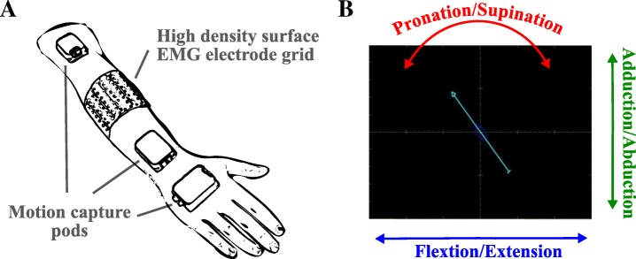
Fig. 1.
The experimental setup (a) and the visual cue provided to the subjects (b). Both the high-density EMG electrodes and the motion capture equipment were fixed with elastic bands to prevent displacements. The position and orientation of the pods were used to calculate wrist joint angles. The retrieved wrist trajectories were stored and later used as labels for training and testing of the estimators. Moreover, the current wrist orientation was directly fed back to the participants in order to support them in executing the cued tasks. Changes in wrist joint angles were reflected in the changes in arrow position and orientation, as seen in panel (b)

