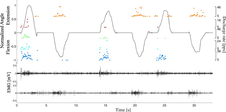
Fig. 5.
Representative example of EMG decomposition during voluntary contractions. Only two EMG channels are shown for clarity (lower traces). The recorded wrist flexion/extension angle is shown in black (upper trace), and a representative subset of decomposed spike trains is represented as dots, whose values indicate instantaneous discharge rates (right axes). The full automatic decomposition introduced errors in spike identification, including missed spiking activity (e.g., third extension). In this example, only one DoF is depicted for clarity and the steady kinematic output during rests between motions is a result of sensors’ intrinsic inertial properties [43]

