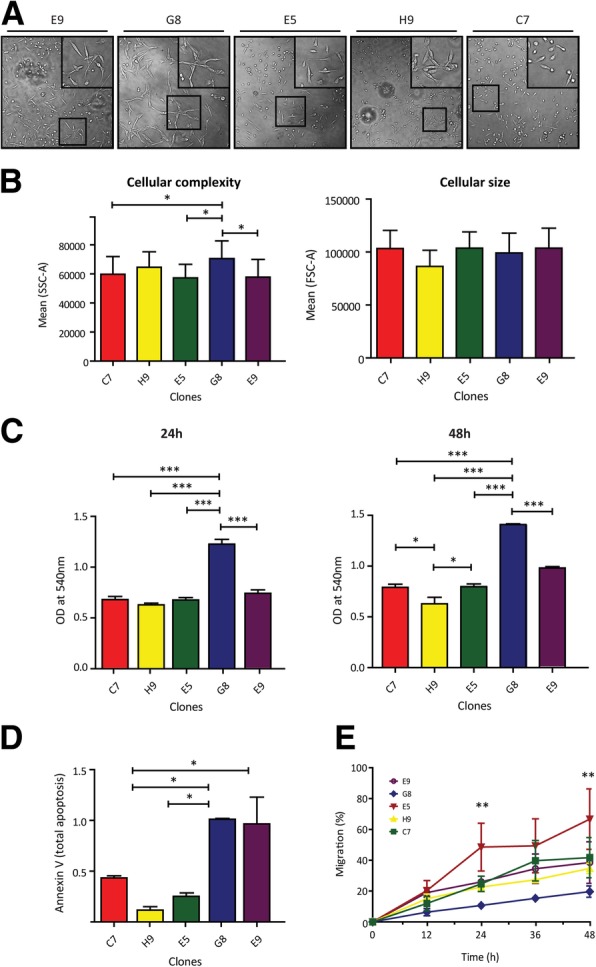
Fig. 3.

Morphology and behavior features of cellular clones derived from MDA-MB 231 cancer cell line. a. Optical microscopy images showing the growth pattern of different MDA-MB 231 derived clones. While C7, E5 and E9 clones presented fibroblast-like phenotype (isolated cell grown pattern), H9 and G8 clones presented epithelial like phenotype (grouped cell pattern) (figure insets). b. As can be observed in flow cytometry analysis, the clones present significant differences in cellular complexity and not in cellular size (SSC and FSC respectively) (ANOVA with Tukey post hoc test p < 0.0016). c. Bar graph showing significant differences in the proliferation rate of the different clones after 24 h by MTT assay (left graph). The differences are increased at 48 h (right graph) (ANOVA with Tukey post hoc test p < 0.0001). d. Bar graph describes significant differences of apoptosis response after 30 min of UV exposure (Anexin V/PI test) (ANOVA with Tukey post hoc test p < 0.0173). e. Migration of different clones was tested by wound-healing assay at 12, 24, 36 and 48 h. As can be observed, clones presented significant differences at 24 and 48 h of migration (ANOVA pos-hoc Tukey test P < 0.0021). Asterisks in all panels indicate significant differences (*, p < 0.05; **, p < 0.01; ***, p < 0.0001)
