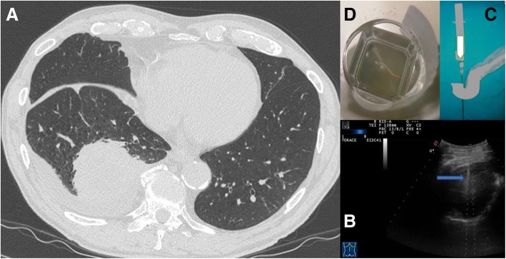
Fig. 1.
a Axial chest computed tomographic (CT) image detected a solid lung nodule suggestive of malignancy in the periphery of the right middle lobe. This lesion has broad pleural contact. b The same lung nodule seen during Ultrasound guided biopsy. We can see the needle (blue arrow) within the consolidation in the right middle lobe.c A dedicated probe with a central hole through which the needle set is introduced. d Specimen suitable for histologic diagnosis (adenocarcinoma)

