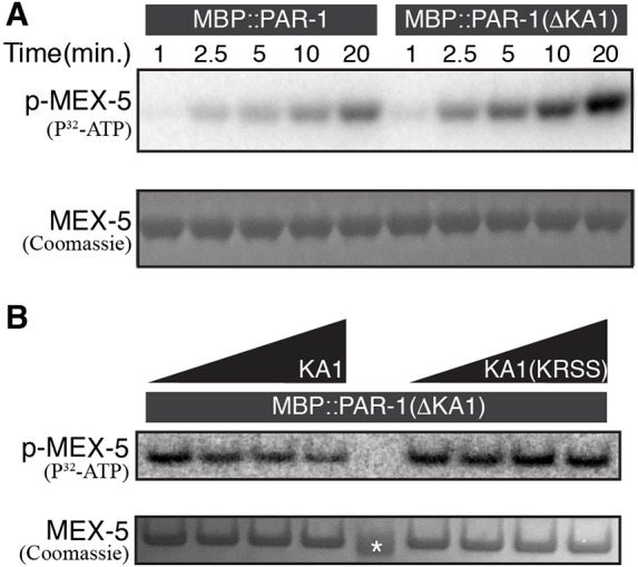
Fig. 4.

The KA1 domain inhibits PAR-1 kinase activity in vitro. (A) Autoradiograph of a time course kinase assay using recombinant MBP::PAR-1 or MBP::PAR-1(ΔKA1) kinase, and MBP::MEX-5(452-460) as a substrate. Reactions were performed in the presence of P32-ATP for the times indicated. Bottom panel shows Coomassie Blue staining to control for loading of MEX-5. The experiment was repeated three times; a representative gel is shown. For loading control of kinase refer to full gels shown in Fig. S4C. (B) Autoradiograph of a kinase assay using recombinant MBP::PAR-1(ΔKA1) kinase and titrating in His(6)::KA1 or His(6)::KA1(KRSS) at 0, 5 mM, 20 mM and 50 mM concentrations (left to right). Reactions were performed in the presence of P32-ATP for 10 min. The experiment was repeated three times; a representative gel is shown. Bottom panel shows Coomassie Blue staining to control for loading of MEX-5. Asterix indicates 37 kDa molecular mass marker. For loading control of kinase refer to full gel shown in Fig. S4D.
