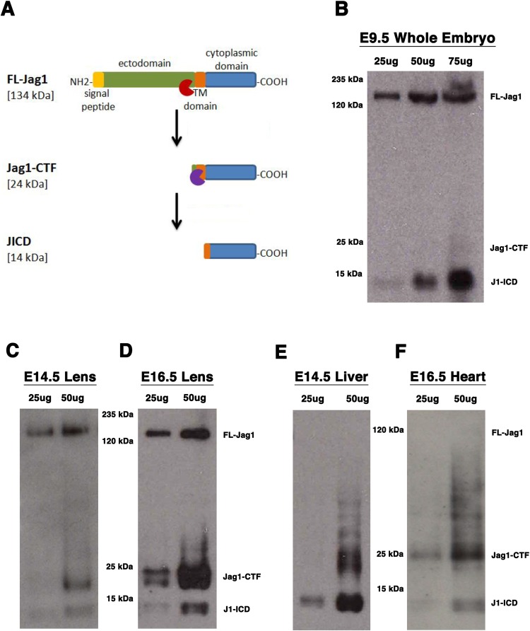
Fig. 1.
Survey of mouse Jagged1 protein isoforms in multiple embryonic tissues. (A) Schematic of Jag1 processed isoforms and predicted molecular weights. Red and purple symbols represent the presumed cleavages by ADAM (red) or γ-secretase (purple) activities. (B–F) Western blot analysis using a C-terminal specific Jag1 antibodies that recognizes all protein isoforms in rodent and human cells. (B) Whole E9.5 mouse embryo extract containing FL-Jag1, Jag1-CTF (seen in longer exposure), and J1-ICD, using goat anti-Jag1 antibody. All three Jag1 isoforms are also detected in the developing lens at (C) E14.5 and (D) E16.5. The only detectable isoforms in (E) E14.5 liver and (F) E16.5 heart tissues are the Jag1-CTF and J1-ICD. Panels C–F were generated using rabbit anti-Jag1 antibody. All blots are representative of three independent protein preparations and western blots (biological replicates).

