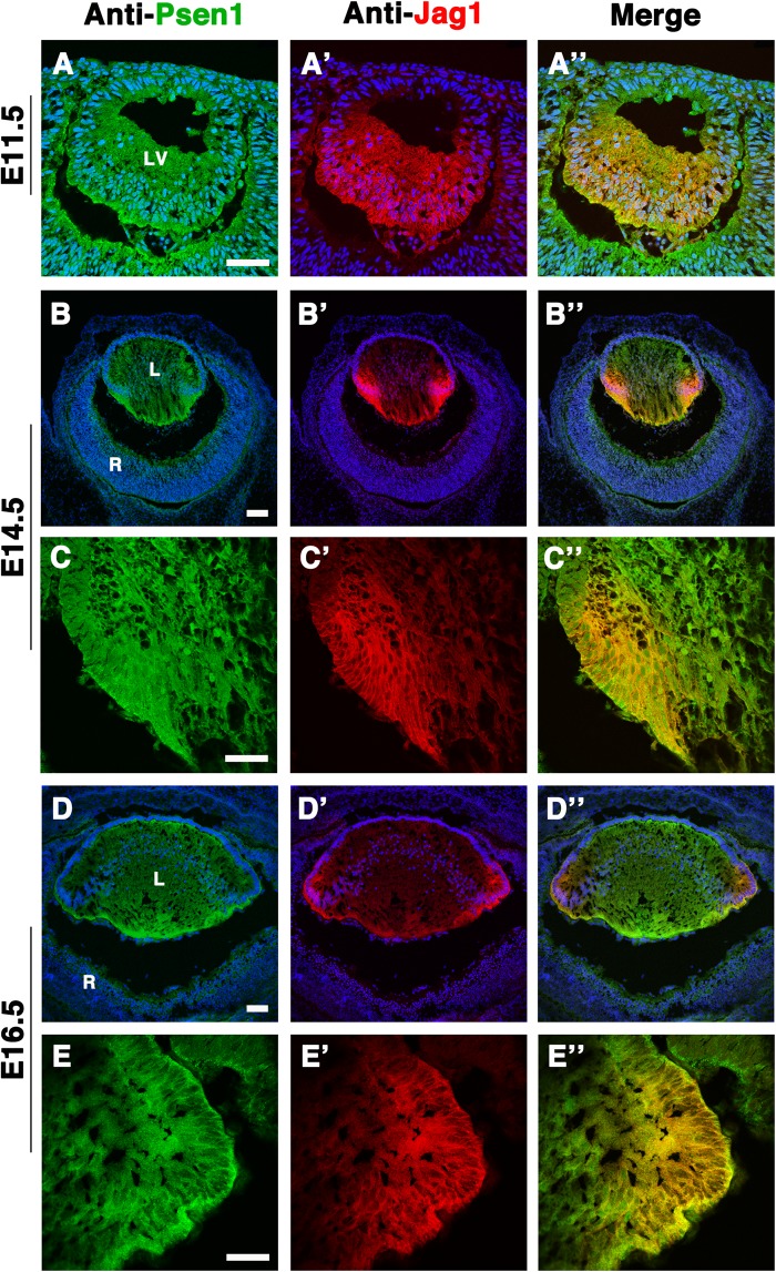
Fig. 2.
Jag1 and Psen1 co-localization in the prenatal mouse lens. Double antibody labelling of wild-type E11.5 lens cryosections (A–A″) demonstrating that Psen1, being ubiquitously expressed in the developing lens (Azimi et al., 2018), has overlap with the Jag1 protein domain (visualized using goat anti-Jag1) that marks the posterior half of the lens vesicle, where cells are beginning to differentiate into primary fiber cells. At E14.5 (B–B″) and E16.5 (D–D″) Jag1 and Psen1 colocalization becomes confined to the Jag1-expressing domain that is now restricted at the lens transition zone, seen more clearly in close-up images (C–C″) and (E–E″), respectively. n=4 biological replicates at E11.5 and E14.5; n=2 biological replicates at E16.5. Anterior is up in all panels. LV, lens vesicle; L, lens; R, retina. Scale bar: in A,C,E = 50 µm, in B,D = 100 µm.

