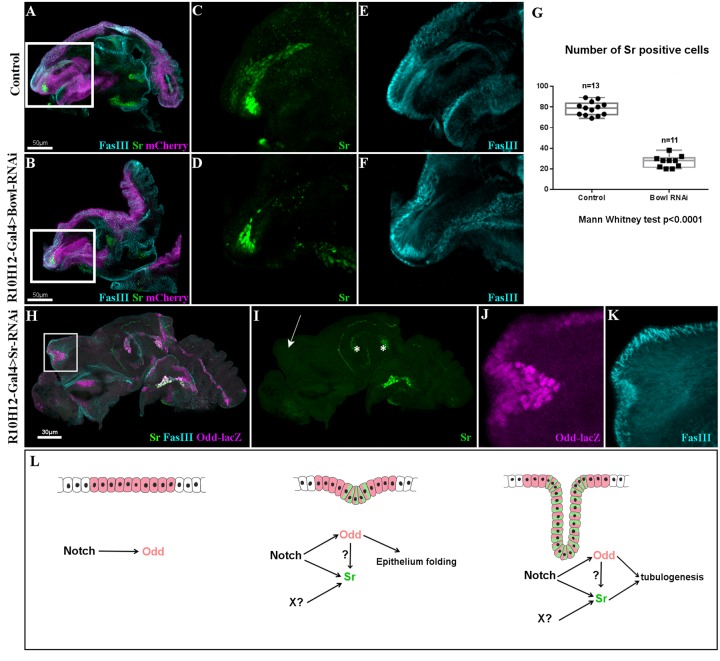
Fig. 5.
Odd-skipped and Stripe interact to form long internal tendons. (A–F) R10H12-Gal4>UAS-mCherryCAAX (magenta) leg discs at 5 h APF immunostained with anti-Sr (green) and anti-FasIII (cyan). (A) Control leg disc and (B) leg disc expressing UAS-BowlRNAi. (C,E) Higher magnifications from A showing Sr-expressing cells forming a long internal tube that elongates into the dorsal femur cavity. (D,F) Higher magnifications from B; number of cells expressing Sr is significantly reduced after Bowl-RNAi expression (D), remaining Sr-positive cells can still invaginate to form a tube reduced in size (compare F with E). (G) Box-plot diagram comparing number of Sr-positive cells in dorsal femur tendon in control and after expression of UAS-BowlRNAi. (H) R10H12-Gal4;Odd-lacZ>UAS-SrRNAi leg discs at 5 h APF immunostained with anti-lacZ (magenta) and anti-FasIII (cyan) and anti-Sr (green). (I) Single channel showing complete absence of Sr protein in dorsal femur where R10H12-Gal4 drives UAS-SrRNAi expression (arrow). Conversely, Sr is still expressed in other tendons (asterisks). (J,K) High magnification of dorsal femur from (H) for Odd-lacZ and FasIII channels respectively. Odd-lacZ expression is maintained in absence of Sr, FasIII accumulation at disc surface indicates that Odd-lacZ+ cells can trigger constriction of the disc epithelium but cannot form an internal tube. (M) In our model, Notch induces Odd in presumptive true joints leading to epithelium folding, Notch/Odd conjointly with unknown signal (X) trigger Sr expression responsible for tendon elongation.

