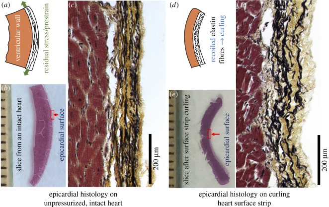
Figure 7.
A histological study to compare the epicardial microstructure on the unpressurized intact heart to the epicardial microstructure on the curled heart surface strip. Movat's pentachrome staining was performed, in which elastin was black, collagen yellow and muscle red. (a) Schematic illustration shows the existence of prestraining/residual stress. (b) Histological slice from an intact heart. (c) On the intact heart, elastin network in epicardium appears less wavy, with a pre-tensioned morphology. (d) Schematic illustration shows that the recoiled elastin fibres bend the bi-layered surface strip. (e) Histological slice obtained after full curling of the native heart surface strip. (f) On the curled surface strip, the elastin pre-tensioned morphology disappears and the elastin network appears recoiled and having more waviness, indicating the release of residual stress. (Online version in colour.)

