Fig. 1.
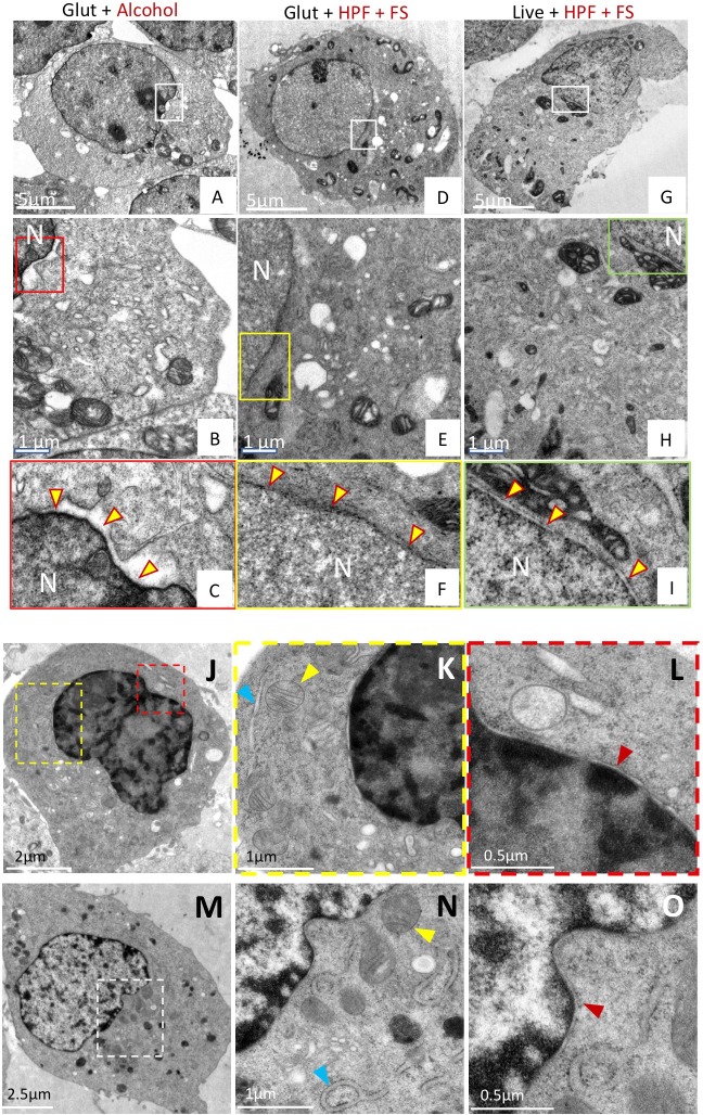
A combination of chemical fixation and cryofixation exhibits superior ultrastructure preservation and membrane staining over traditional methods in HEK-293T cells. (A–I) HEK-293T cells. Cells prepared by conventional glutaraldehyde fixation and dehydration methods (A–C), glutaraldehyde followed by cryofixation (D–F) or cryofixation alone (G–I) were examined by thin-section EM. Panels B,E,H represent magnified views from boxed regions of panels A,D,G, respectively. Panels C,F,I represent magnified views of the boxed regions from panels B,E,H, respectively, and highlight the preservation of the nuclear membrane in each case (yellow arrowheads). The nuclear membrane appears smooth and uncompromised in samples prepared by glutaraldehyde treatment and cryofixation (Glut+HPF+FS) or cryofixation alone (Live+HPF+FS), but is ruffled and irregular in samples prepared by conventional means (Glut+Alcohol). (J–O) BHK cells. Ultrastructural preservation in cells prepared by the combined (Glut+HPF+FS) method (M–O) was comparable to that in cryofixed (Live+HPF+FS) cells (J–L), as illustrated by the presence of intact mitochondria (yellow arrowheads, panels K and N), intact ER (blue arrowheads, panels K and N), and absence of ruffled nuclear membranes (red arrowheads, panels L and O). Panels K and L are magnified views of the respective yellow and red boxes in panel J. Panel O is a magnified view of panel N, which is a magnified view of the region delineated by the white box in panel M. Glut, glutaraldehyde; HPF, high-pressure freezing; FS, freeze substitution; N, nucleus.

