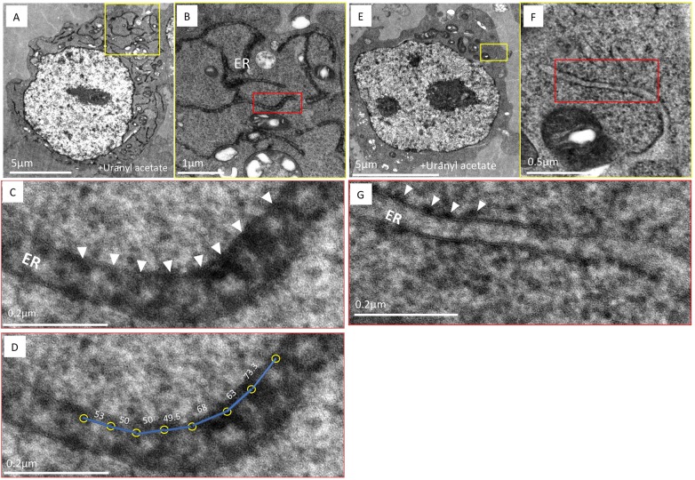
Fig. 3.
HYPE localizes to the lumenal face of the ER membrane as periodic foci. (A) An image of a thin section of HEK-293T cells expressing HYPE–APEX2 and processed by cryoAPEX reveal staining of the ER tubules in a well-preserved (dense) cytoplasmic background. (B,C) Higher magnification images of a small section of the peripheral ER (demarcated by yellow box in A and shown in B, with further magnification of red box in B shown in C) exhibits periodic foci of APEX2-generated density (B, red box and C, white arrowheads showing periodicity between the HYPE foci). (D) Center-to-center density measurements showing the distance (in nm, blue lines) between the HYPE-specific foci (yellow circles) in C. (E) Untransfected control HEK-293T cells processed in an identical manner show the lack of APEX2-generated density within the ER lumen. (F,G) Higher magnification images of a small section of E (demarcated by yellow box and shown in F, with further magnification of red box in F shown in G) clearly shows the lack of density on the lumenal face. Additionally, density corresponding to ribosomes on the cytoplasmic face of the ER membrane is evident (G, white arrowheads). Thus, HYPE is detected only along the lumenal face of the ER membrane and never on the cytosolic face.

