Fig. 5.
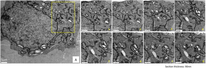
Superior ultrastructural preservation enables the tracking of HYPE's subcellular localization via serial sectioning. To demonstrate the consistency of the membrane ultrastructure preservation obtained by cryoAPEX, cells expressing HYPE–APEX2 were serially sectioned and a specific area (panel A, yellow box) was imaged. Multiple ribbons containing between 10 and 20 serial sections of 90 nm thickness were collected, screened and imaged. Representative images of eight serial sections showing ER localization of HYPE are presented (images serially numbered 1–8). Sections exhibit a dense well-preserved cytoplasm with undisrupted membrane ultrastructure of organelles such as mitochondria and Golgi complex in close proximity to the ER tubules containing HYPE–APEX2 density. Thus, we can follow HYPE localization through the volume of the cell without loss of contextual information. Scale bars: 5 μm (A); 8 μm (magnifcations 1–8).

