Fig. 2.
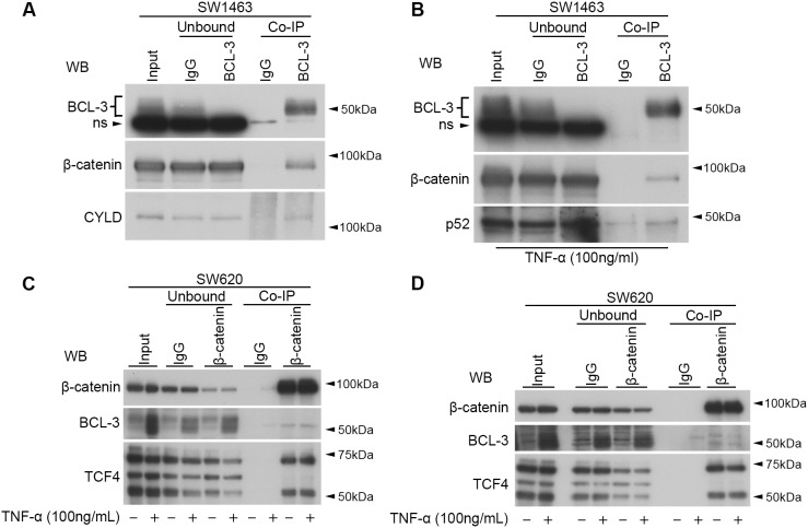
BCL-3 interacts with β-catenin in CRC cell lines. (A,B) BCL-3 Co-IP performed in SW1463 nuclear-enriched lysates. Unbound (immuno-depleted) lysates show depletion of proteins after IP. Immunoprecipitates were analysed by western blot for BCL-3 and β-catenin. IgG serves as a negative control. (A) CYLD serves as a positive control for BCL-3 binding. (B) Cells were treated with 100 ng/ml TNF-α for 6 h prior to lysis. Immunoprecipitates were additionally analysed for p52. Note the presence of a non-specific (ns) band in BCL-3 western analysis with use of Abcam anti-BCL-3 antibody. (C,D) Nuclear β-catenin Co-IPs in SW620 cells. Panels C and D represent experimental replicates. Western analysis of non-treated and 6-h TNF-α-treated cells following β-catenin Co-IP. TCF4 serves as a positive control for β-catenin binding. Mouse pan-IgG serves as a negative control. Unbound, immuno-depleted lysates following Co-IP; WB, western blot antibody.

