Figure 5.

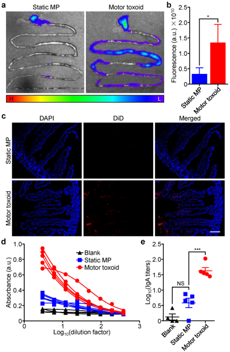
In vivo delivery and antibody titer generation. (a) Representative images of the gastrointestinal tract of male CD-1 mice 6 h after administration of DiD-labeled static microparticles (MP) or motor toxoids by oral gavage (H: high fluorescence, L: low fluorescence). (b) Quantification of the fluorescence from (a) (n = 3, mean + SD). (c) Histological sections from the intestine 6 h after administration of DiD-labeled (red) static MP or motor toxoids; DAPI (blue) was used to label cell nuclei (scale bar = 100 μm). (d) Absorbance data from an ELISA assay probing for IgA antibody production against staphylococcal α-toxin in the feces of mice one week after the administration of blank solution, static MP, or motor toxoids (n = 4 or 5; four-parameter dose-response curve). (e) IgA titers against α-toxin as calculated using the data from (d) (n = 4 to 5, geometric mean ± SEM). *p < 0.05, ***p < 0.001, NS: not significant; one-way ANOVA.
