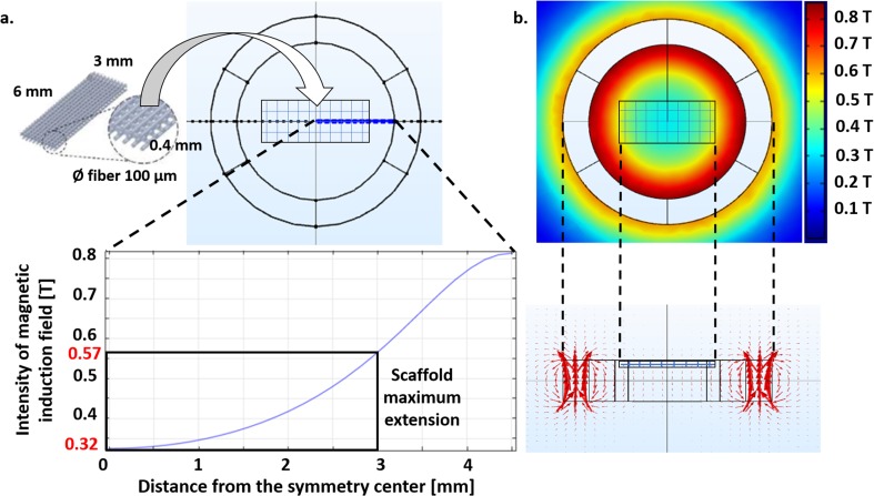
Fig. 3.
Axial-symmetric model of the magnetic field generated in a single culture chamber. a Intensity of magnetic induction field with the distance from the symmetry center of the magnets (blue line). The black rectangle highlights the dimensions of polystyrene scaffolds (6x3x0.4 mm) identical to those for cell experiments. They are obtained by fused deposition modeling and composed of four layers of fibers (diameter: 100 μm, with a pore size: 300 μm) shifted of 150 μm with respect to the adjacent. A sketch of the positioning of the scaffolds in the culture chambers is also shown. b Color map showing the distribution of the magnetic induction field around the magnets (top view) and side view of the arrow field. A sketch of the positioning of the scaffolds in the culture chambers is reported

