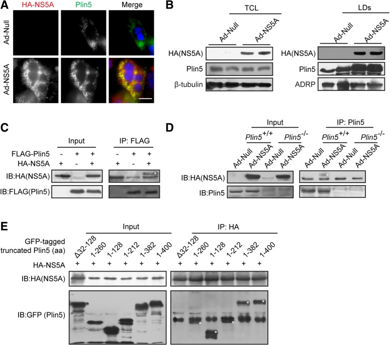
Fig. 5.
NS5A recruited Plin5 to LDs. a NS5A was expressed in primary hepatocytes, and exogenous HA-NS5A (red) and endogenous Plin5 (green) were observed by immunofluorescent staining. Scale bar = 20 μm. b LD-containing fractions were isolated from wild-type mice with or without NS5A expression by using sucrose density gradient ultracentrifugation, and the Plin5 content was determined in both the total cell lysate (TCL) and the lipid droplet (LD)-containing fraction by immunoblotting. c Co-IP assay was performed to determine the interaction of exogenous HA-tagged NS5A and FLAG-tagged Plin5 in 293 T cells. Cell lysates were subjected to immunoprecipitation with anti-FLAG antibody and then immunoblotted with an anti-HA antibody. d NS5A was expressed in wild-type (Plin5+/+) and Plin5-null (Plin5−/−) primary hepatocytes, endogenous Plin5 was immunoprecipitated with anti-Plin5 antibody, and NS5A was detected by immunoblotting with anti-HA antibody. e Co-IP assays to identify the specific domain of Plin5 responsible for the Plin5-NS5A interaction. Truncated GFP-tagged Plin5 constructs were overexpressed in 293 T cells with HA-tagged NS5A. NS5A was immunoprecipitated by anti-HA monoclonal antibody, and truncated Plin5 was detected by immunoblotting

