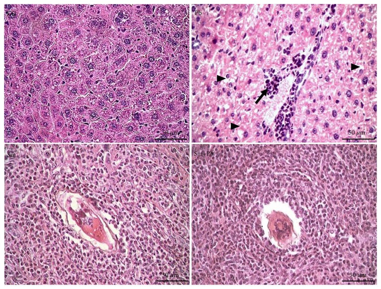
Figure 2.
Microscopic structure of the liver tissue from control mice and those acutely infected with Schistosoma mansoni and Paracoccidioides brasiliensis (Hematoxylin and Eosin staining, ×400 magnification, bars= 50 μm). CG (control group): uninfected mice. Pb: group infected only with P. brasiliensis. Sm: group infected only with S. mansoni. Sm+Pb: group coinfected with S. mansoni and P. brasiliensis. A S. mansoni egg is centrally observed in granulomas. Arrow: perivascular inflammatory infiltrate. Arrowhead: hepatocytes with hydropic degeneration.

