Figure 2.
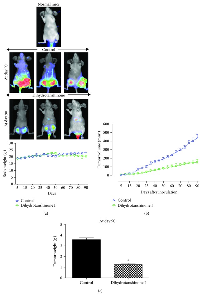
The effect of DHTS on tumor growth in a xenograft model. Three million cells were injected into the mammary fat pad of each immunodeficient NOD-SCID female nude mouse. Analysis of the effect of tumor growth on DHTS and MCF-7 cell-bearing immunodeficient nude mice. The dose of drug used was 10 mg/kg once a week. After 13 weeks, images were captured with an Odyssey® Imager (LICOR, Pearl Image System, USA). The weights of mice were comparable with the control and DHTS-treated groups (a). The high grade of the tumors was detected using the IRDye 800CW optical probe (2DG) in the 800 nm channel, represented in pseudo-color. Tumor volume was measured twice/week using a caliper and calculated as (width2 × length)/2. Tumor growth curves were monitored during the experimental period (b). The effect of DHTS on tumor weights. Tumor weights were measured after therapy. ∗P < 0.05 compared to the control (c). Representative images were captured at the end of 13 weeks of therapy, and the results are shown for vehicle-treated control and DHTS-treated mice.

