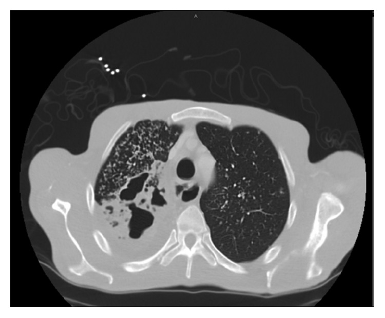
Figure 2.

Contrast-enhanced CT scan. Axial section (neck) showing airspace consolidations with diffuse tree in bud opacities in the right lung apex and to a less extent, in the left lung apex. In addition, there are multiple areas of cavities within the right lung apex.
