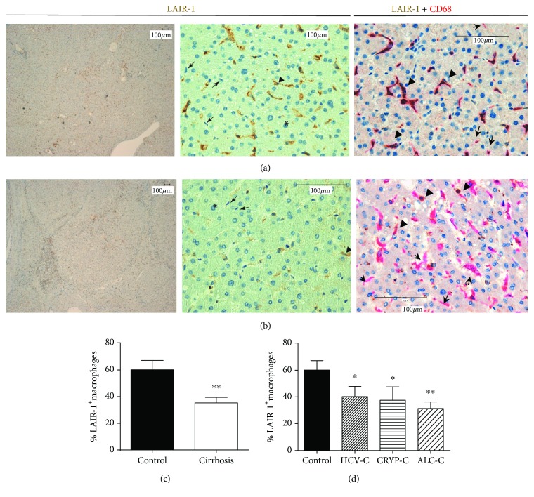
Figure 1.
LAIR-1 expression in cirrhotic human liver. LAIR-1 expression was analyzed by immunohistochemistry in hepatic biopsies from controls (n = 18) (a) and cirrhotic patients (n = 22) (b). Images were obtained using a Leica DM108 with a magnification of 4x (left panels) and 40x (middle and right panels). The asterisks show hepatocytes, the arrowheads show macrophages with positive staining, and the arrows point at macrophages with negative staining for LAIR-1. The number of macrophages (CD68 positive staining in magenta) expressing LAIR-1 (brown) was quantified and compared between control and cirrhosis (c) or between healthy controls and cirrhotic patients attending to their etiology (d). Data represented in histograms are the mean and SEM. Two-sided Mann-Whitney U test: ∗P < 0.05 and ∗∗P < 0.01, between cirrhotic patients and healthy controls.

