Figure 2.
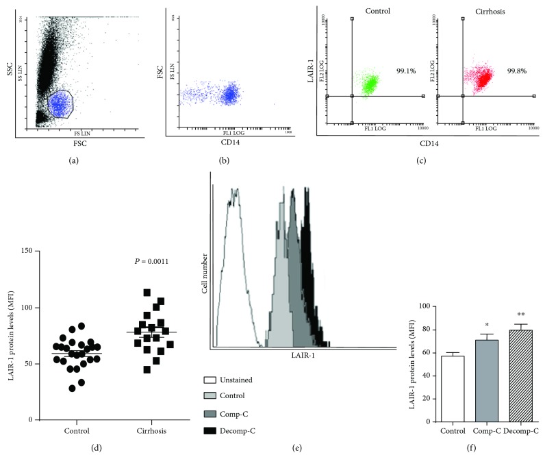
LAIR-1 expression in blood monocytes. LAIR-1 expression was analyzed by flow cytometry in peripheral blood of cirrhotic patients (n = 17) or healthy controls (n = 20). Leukocytes were gated based on forward vs. side scatter (a). Monocytes were gated on the base of both CD14+ cells and morphology (b). Representative dot-plot of monocytes stained with CD14 and LAIR-1 antibodies in healthy controls or cirrhotic patients (c). The cell surface LAIR-1 protein expression was quantified measuring the MFI (median fluorescence intensity) after cell staining and compared between control and cirrhosis (horizontal lines indicate the mean and SEM) (d). Representative histograms of monocytes unstained and stained with LAIR-1 antibody in healthy controls and compensated (Comp-C) or decompensated (Decomp-C) cirrhotic patients are shown (e). The cell surface LAIR-1 protein expression was compared between healthy controls and cirrhotic patients attending to their clinical progression, compensated (Comp-C) or decompensated (Decomp-C) cirrhosis, and represented in histograms (bars represent the mean and SEM) (f). Two-sided Mann-Whitney U test: ∗P < 0.05 and ∗∗P < 0.01, between cirrhotic patients and healthy controls.

