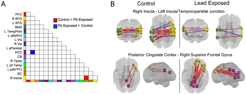
Figure 2. Lead-exposed and lead-naïve participant groups differences in neural connectivity between and across networks.
Differences were observed in two network pairs. Bi-lateral connectivity across insular-temporal cortices showed greater increase with age in lead-naïve fetuses (panel B upper), whereas lead-exposed fetuses showed a strengthening of lateral prefrontal (SFG) to posterior cingulate (PCC) connectivity with age that was not present in the comparison group (panel B lower). Both observations suggest lack of advancement of typical processes with the same rate in lead-exposed fetuses, as cross-hemispheric increases and reduced PCC-SFG have been observed as normative fetal brain developmental processes (Jakab et al., 2014; Thomason et al., 2014).

