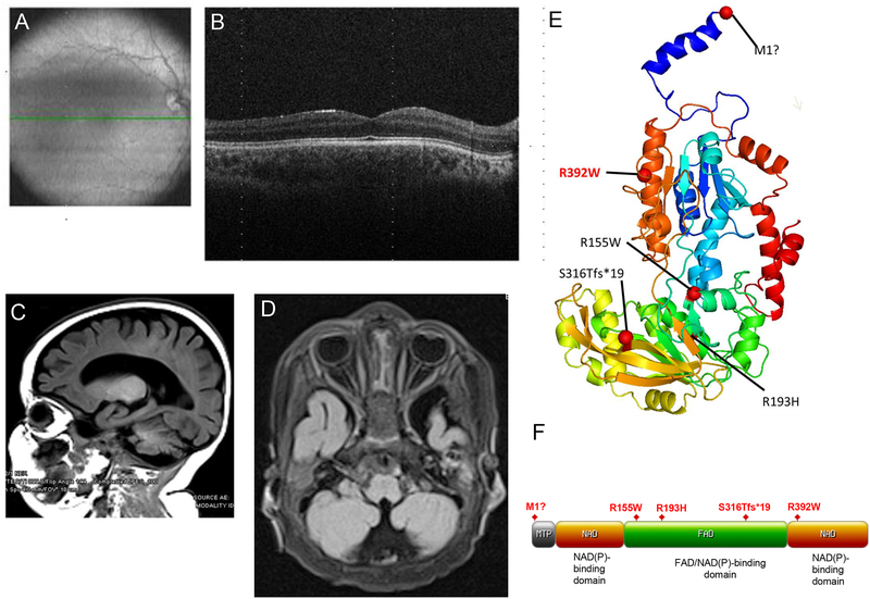
Figure 1. Optic atrophy and cerebral atrophy in patients carrying biallelic mutations in FDXR.
(A–B) Optical coherence tomography (OCT) of retina from the proband of Family #1. Images were obtained at the age of 35 months. The green line shown in the fundus photograph in panel A indicates the position of the OCT scan shown in panel B. The fovea appears to be well-formed, and the outer retinal layers are normal. The inner retina, however, shows almost complete absence of the ganglion cell layer in both eyes and severe thinning of the nerve fiber layer, consistent with bilateral optic atrophy involving the papillomacular bundle. (C–D) MRI images from the proband of Family #2 also show optic nerve hypoplasia (C) and global cerebral atrophy with marked prominence of sulci and ventricles (D). (E) Patient mutations were mapped to a three-dimensional FDXR protein structure model. The most common variant observed in all patient families (R392W) (10) has been highlighted in red for comparison. (F) Patient mutations were also mapped to a two-dimensional map of the FDXR functional domains. Domain map was generated using the “MyDomains” software provided by PROSITE (34).

