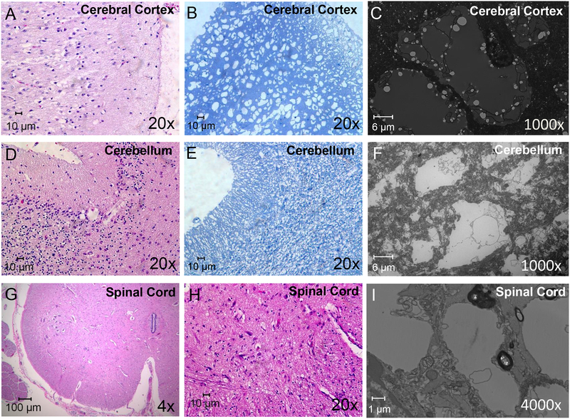
Figure 2. Atrophy and cell loss in the CNS of the patient autopsy (proband 2) with FDXR mutations.
Histological sectioning of the occipital cerebral cortex with H&E staining (A) indicated that the cerebral cortex lost many of its neurons, and multiple vacuoles were observed in the H&E staining section that formed a spongiform structure. Toluidine staining of the cerebral section (B) also showed the presence of spongiform-like structures in the cerebral cortex, and TEM confirmed this was largely due to the presence of vacuole-like structures (C). The cerebellum of the patient also lost neurons in the molecular layer, Purkinje cell layer, and granular layer (D–F). Most of the Purkinje cells were absent, and multiple vacuoles were observed with H&E staining (D). Toluidine staining (E) and TEM imaging (F) of the cerebellar sections also showed loss of cells and the formation of vacuoles in the cerebellum. H&E staining (G, H) of spinal cord also indicated that the spinal cord lost lower motor neurons in the anterior horn (G), and multiple vacuoles were formed in the spinal cord by H&E staining (H) and TEM (I).

