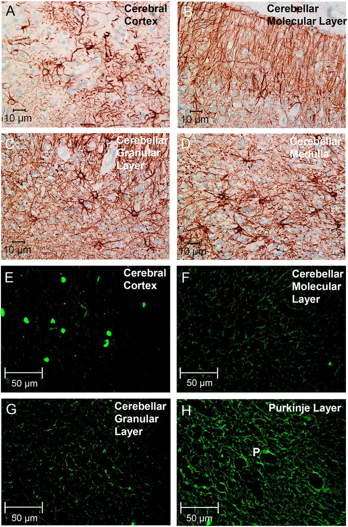
Figure 3. Signs of gliosis and neurodegeneration in the autopsy brain from proband 2.
Immunostaining with antibody specific for GFAP revealed extensive gliosis in the cerebral cortex (A), as well as in the molecular layer (B), granular layer (C), and medullary layer (D) of the cerebellum. Astrocytes and their extending branches were observed throughout the cerebrum and cerebellum, and Bergmann glial cells were prominent throughout the molecular layer (B). Immunostaining for Fluoro-Jade C (FJC) also showed signs of neurodegeneration in the cerebral cortex (E), the cerebellar molecular layer (F), the cerebellar granular layer (G), and in the Purkinje layer (H, Purkinje cell indicated by the letter “P”).

