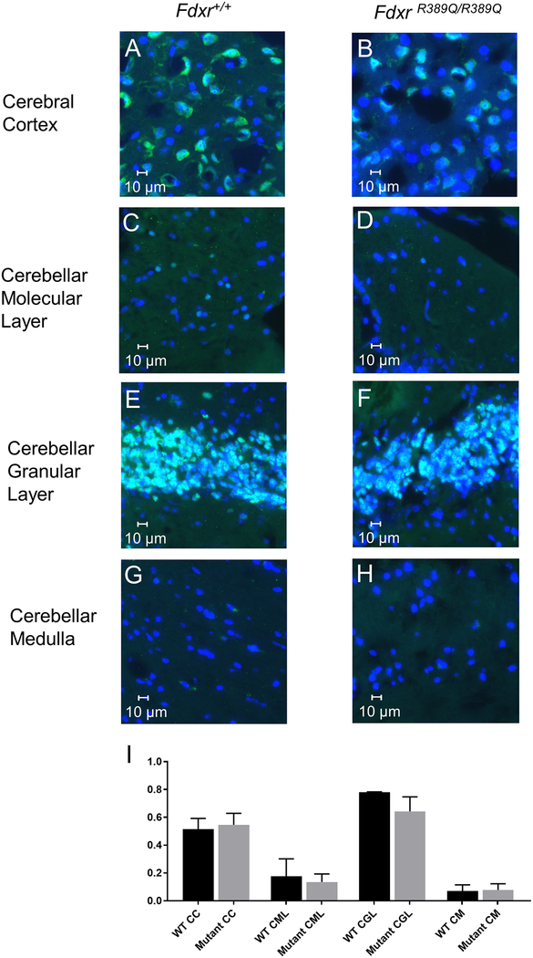
Figure 6. Modest reduction in the proportion of neurons in Fdxr mutant brains.
IF staining with anti-NeuN antibody was used to determine the proportion of neurons in mutant and wildtype brains. Counterstaining with DAPI was used to show the total number of cells in each image field; cells staining with DAPI alone are largely composed of glial cells, with a small percentage consisting of neurons such as Purkinje cells that do not express NeuN. Staining reveals a similar number of neurons present in the cerebral cortex (A–B), molecular layer (C–D), and medullary layer (G–H) of the cerebellum between Fdxr mutant and wildtype mice. The number of neurons present in the granular layer of mutant cerebellar tissue (E–F) is modestly, but noticeably reduced relative to wildtype control tissues. The proportion of neurons in each brain region and genotype was also determined by dividing the number of NeuN-positive cells by the total number of DAPI-positive cells (I), confirming the modest reduction in neurons seen in the granular layer. Cell counts were generated by manual counting of cells in 3 different fields of view at 40X magnification, and then averaging the result from the 3 counts. A total of 174 to 834 cells were counted for each genotype and brain region / layer, which was determined by the density of neurons in each brain region. Abbreviations are as follows: “CC” = Cerebral Cortex, “CML” = Cerebellar Molecular Layer, “CGL” = Cerebellar Granular Layer, “CM” = Cerebellar Medulla, “WT” = Wildtype.

