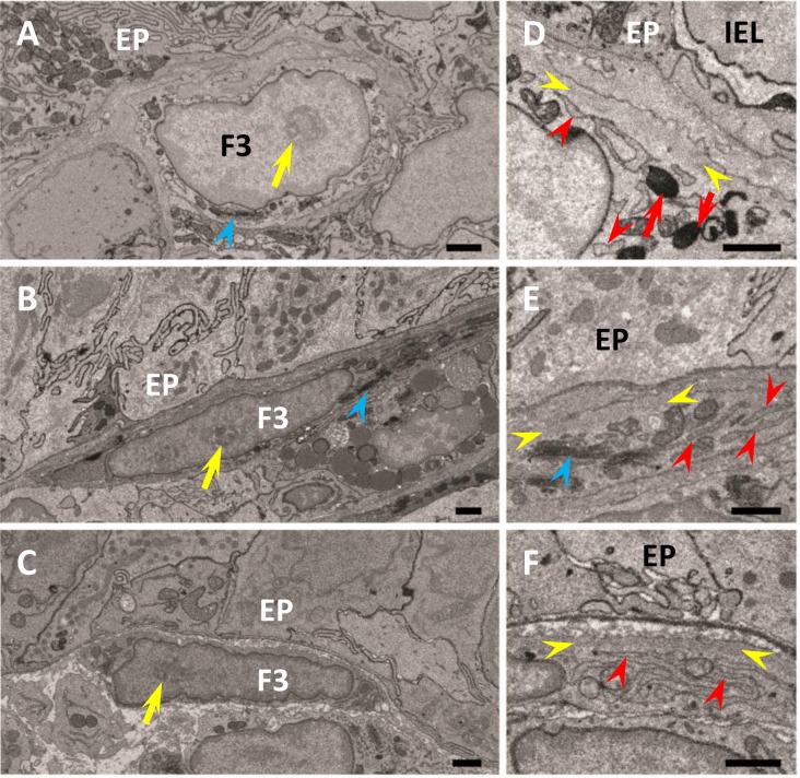
Fig. 4.
Ultrastructure of FBLC type III in the apical (A, D) and basal (B, E) portions of the intestinal villus and around the lateral portion of the intestinal crypt (C, F). FBLC type III in all portions possess a nucleolus (yellow arrows) and Golgi apparatus (blue arrowheads). A non-organelle region with high electron density is visible in FBLC type III in the apical (D) and basal (E) portions of the intestinal villus and around the intestinal crypt (F) (region between yellow arrowheads). FBLC type III in the apical portion of the intestinal villus possess several lysosomes (red arrows). Endoplasmic reticula in FBLC type III in the basal portion of the intestinal villus and around the intestinal crypt show a thin lamellar shape (E, F, red arrowheads), while those in the apical portion of the intestinal villus are irregularly arranged and slightly expanded (D, red arrowheads). EP, epithelium. IEL, intraepithelial lymphocyte. F3, FBLC type III. Bar=1 µm.

