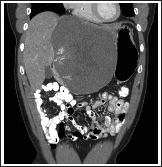
Figure 1.

12cm × 14cm low density heterogeneous mass within the left hepatic lobe on CT performed in the Fall of 2010. The mass demonstrated peripheral pooling of contrast initially with centripetal filling on delayed images using triphase IV contrast abdominal CT, consistent with a cavernous hemangioma.
