Figure 2.

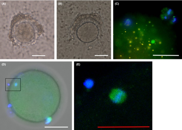
Collected oocytes from resected ovaries and immunofluorescent staining. A, Collected immature oocytes before in vitro maturation in patient 8. B, An oocyte in meiosis II showing the first polar body after in vitro maturation in patient 8. C, Immunofluorescent staining of a mature oocyte in patient 5. D, Immunofluorescent staining of a mature oocyte in patient 13. The spindle is enclosed in a square box. E, Enlarged view of the spindle corresponding to the boxed area in (D). White scale bar indicates 50 µm. Red scale bar indicates 25 µm. Green, microtubules; blue, DAPI
