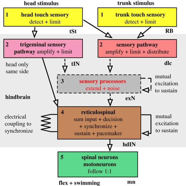
Figure 2.

Organization of neuronal pathways from head and trunk skin stimulation to swimming in the hatchling frog tadpole. The boxes represent populations of similar excitatory neurons in the five functional levels between a skin stimulus and a motor response. Arrows show direct excitatory synaptic connections established by recording from pre- and post-synaptic neurons. Dashed boxes and arrows are populations and connections proposed to explain current evidence. For simplicity, the pathways are only shown for one side of the body and inhibitory neurons are omitted. Letters near boxes are the abbreviations of neuron names they contain. (Online version in colour.)
