Figure 3.
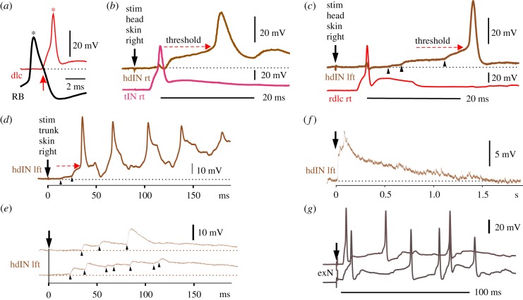
Recordings show the activity and connections of neurons in the swimming response pathway: (a) current injection into a sensory RB neuron leads to an action potential (*) and direct excitation of a sensory pathway dlc neuron (excitatory post-synaptic potential (EPSP) starts at arrow) which then evokes an action potential in the dlc (*). (b,c) Paired recordings showing responses to a 1 ms head skin current stimulus which evoked swimming. (b) A sensory pathway tIN responds with a single spike. The excitation from this and other tIN spikes excites the hdIN on the same side which depolarizes smoothly to threshold (dashed arrow) and fires a spike. (c) A rostral dlc on the right side also fires a single spike but this cannot explain the slow, noisy build-up of excitation (arrowheads) to threshold (dashed arrow) and firing in the hdIN on the opposite side. (d–f) Trunk skin stimulation also leads to variable excitation of hdINs. (d) A stronger stimulus leads to two jumps (arrowheads) in hdIN which reach threshold (arrow) and firing is followed by rhythmic firing during swimming. (e) Two examples of responses subthreshold for swimming show noisy excitation (arrowheads) summing but not reaching firing threshold. (f) The long duration of subthreshold excitation in hdINs is revealed by averaging five recordings at a slower time-scale. (g) Two examples of responses of a possible sensory processing neuron (exN) in the hindbrain to a trunk skin stimulus (at arrow). Each record shows an early spike and variable later firing. (Online version in colour.)

