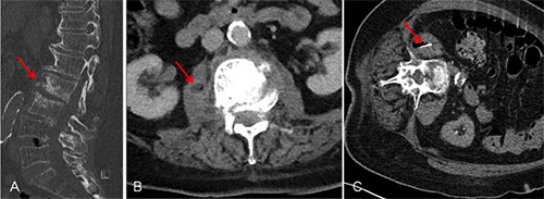
Figure 1.

Computed tomography (CT) guided biopsy of paravertebral soft tissue in a patient with clinical and radiological signs of spondylodiscitis. A) Sagittal reformatted computed tomography in bone window illustrating partially destroyed and eroded vertebral bodies at lumbar segment 2 and 3 (arrow). B) Computed tomography in axial direction showing fluid collection with entrapped air in the right iliopsoas muscle (arrow). C) Tip of the biopsy needle (arrow) located in the paravertebral soft tissue (right ilipsoas muscle).
