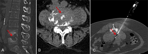
Figure 2.

Computed tomography (CT) guided biopsy of intervertebral disc in a patient with clinical and radiological signs of spondylodiscitis at lumbar segment four and five. A) Sagittal reformatted computed tomography in bone window showing destruction of intervertebral disc and partially destroyed vertebral bodies (arrow). B) Transversal computed tomography illustrating gross inflammatory changes in lumbar segment 4/5 (arrow). C) Transversal computed tomography in bone window showing biopsy needle with co-axial technique targeting intervertebral disc (arrow).
