
Figure 1.
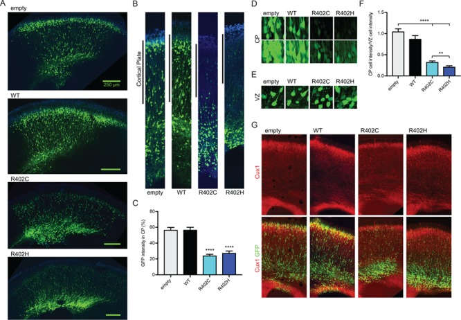
Tuba1a-R402C/H mutants dominantly disrupt neuronal migration in the developing mouse cortex. (A) Coronal sections from E18.5 mouse brain electroporated at E14.5 with pCIG2 vectors: empty vector (empty), WT TUBA1A (WT), TUBA1A-R402C (R402C) or TUBA1A-R402H (R402H). (B) Representative regions of cortex analyzed for cortical plate fluorescence. (C) Percentage of GFP signal in the cortical plate. For each condition, three coronal sections from at least four separate animals were analyzed. Data are represented as mean ± SEM. Quadruple asterisks indicate significant difference compared to WT, by t-test (P < 0.0001). (D) Representative images of cortical plate neurons for each condition, at two different exposures. (E) Representative images ventricular zone neurons for each condition. (F) Quantification of mean GFP intensity of cortical plate neurons normalized GFP intensity of ventricular zone neurons. Data are represented as mean ± SEM. At least 45 neurons for each condition were measured (Region of interest (ROI) = 200 μm2). Quadruple asterisks indicate significant difference compared to WT, by t-test (P < 0.0001). Double asterisks indicate significant difference compared to WT, by t-test (P < 0.01). (G) Cux1 staining of electroporated sections reveals position of upper layer cortical neurons.