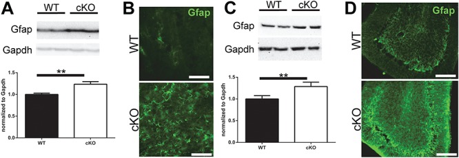
Figure 7.

Reactive gliosis in Thap1 cKO mouse striatal and cerebellar tissue. Western blot protein expression and quantification of Gfap in (A) striatal and (C) cerebellar tissue (n = 7, paired t-test; P < 0.01). Representative images of Gfap immunostaining in STR (B) and CB (C) of Thap1 cKO and wild-type mice at 4 months (scale bar: 50 μm).
