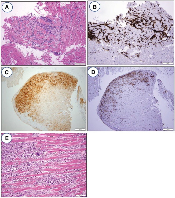
Figure 3.
Histology and immunohistochemistry of ICB myocarditis. (A) Photomicrograph of endomyocardial biopsy showing lymphocytic myocarditis with a dense mononuclear cell infiltrate, associated myocyte damage, and oedema. H&E stained section, ×200 original magnification. (B) Immunohistochemistry for CD3 demonstrating that the infiltrate is predominantly composed of T lymphocytes, ×200 original magnification. (C) Immunohistochemistry for PD-L1 shows positive staining of myocytes only in the areas of lymphocytic myocarditis, ×100 original magnification. (D) Immunohistochemistry for HLA-DR, a surrogate marker for IFN-γ activity, shows positive staining in the areas of lymphocytic myocarditis, ×100 original magnification. (E) Photomicrograph of endomyocardial biopsy showing giant cell myocarditis with an inflammatory infiltrate, extensive myocyte damage, and the presence of foreign body giant cells. This type of myocarditis can be seen in patients on ICB therapy. H&E stained section, ×200 original magnification.

