Figure 5.
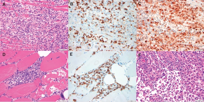
Histologic findings from ICI-associated myocarditis. (A–C) Photomicrographs of the myocardium, specifically, the interventricular septum. (A) Dense mononuclear cell infiltrate with extensive myocyte damage and necrosis, consistent with lymphocytic myocarditis, H&E stained section. (B) Abundance of CD3-positive T cells, CD3 immunohistochemical stain. (C) Prominent CD68-positive macrophage infiltrate, CD68 immunohistochemical stain. (D–E) are photomicrographs of skeletal muscle. (D) Dense mononuclear cell infiltrate and evidence of myocyte necrosis and damage with nuclear internalization, demonstrating myositis, H&E stained section. (E) Abundance of CD-3 positive T cells, CD3 immunohistochemical stain. (F) Photomicrograph of metastatic focus of melanoma with a lymphocytic infiltrate, H&E stained section. All histology displayed at 40× original magnification. All scale bars in this figure denote 50 µm. The patient was a 65-year-old woman who received one dose of nivolumab combination therapy with ipilimumab.

