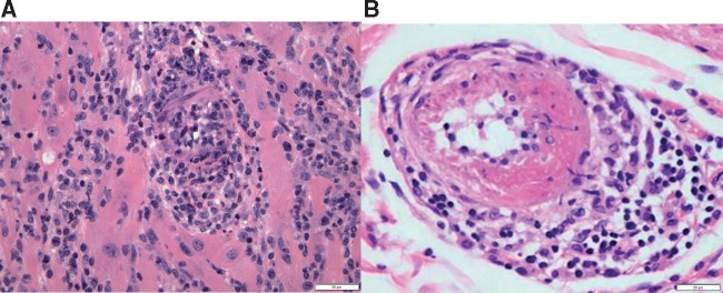
Figure 8.
Histologic findings from ICI-associated vasculitis. (A) Photomicrograph of myocardium with lymphocytic myocarditis and vasculitis, with obliteration of the lumen of this small artery. H&E stained section, 200× original magnification, with scale bar denoting 50 µm. (B) Photomicrograph of small artery with fibrinoid necrosis and perivascular chronic inflammation. H&E stained section, 400× original magnification, with scale bar denoting 20 µm.

