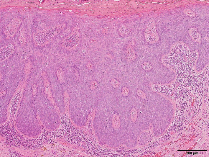
Figure 2.

Histopathology examination showed irregularly acanthotic squamous epithelium with markedly atypical keratinocytes, many mitoses, and a loss of polarity but without invasion, thus confirming the diagnosis of a carcinoma in situ (H&E stain, original magnification ×100)
