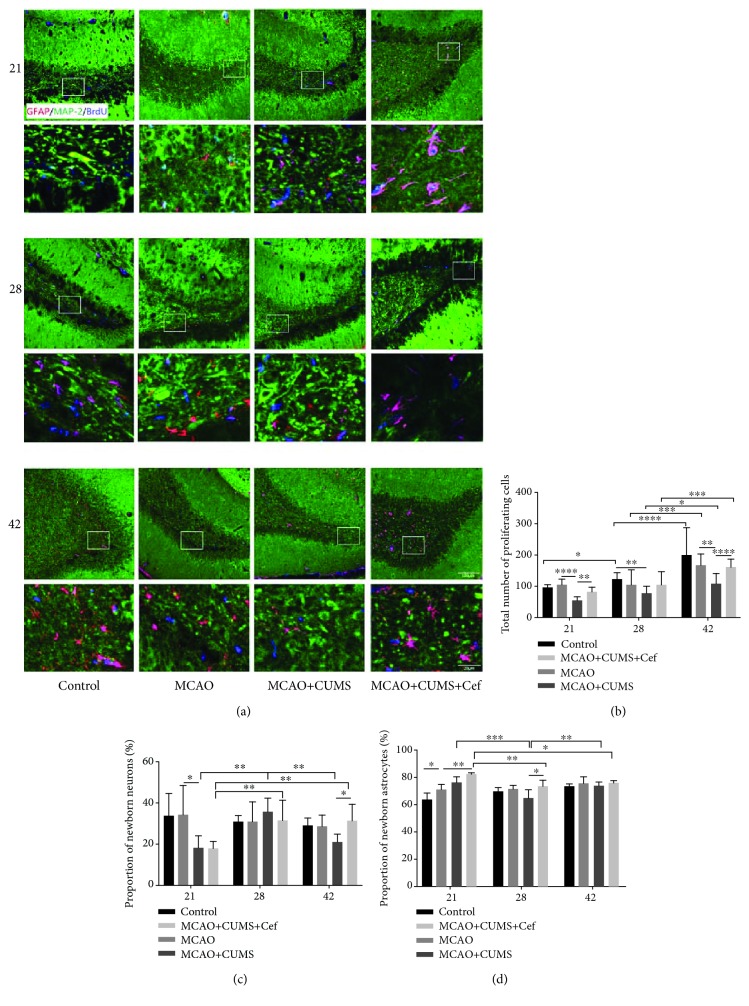
Figure 2.
Ceftriaxone improved regeneration capacity impaired in the hippocampal DG of the CUMS-induced model rats. (a) Representative immunofluorescent photomicrographs (200×) of GFAP/MAP2/BrdU (red/green/blue) in hippocampal DG were pictured on the 21st/28th/42nd day. Scale bars: 100 μm and 20 μm. (b) Ceftriaxone promoted the proliferation of total newborn cells in hippocampal DG of the CUMS-induced model rats. The newborn nuclei were labeled by BrdU (blue). (c) Ceftriaxone influenced the proportion of newborn neurons (MAP2/BrdU, green/blue) in the total newborn cells. (d) Ceftriaxone raised the proportion of newborn astrocytes (GFAP/BrdU, red/blue) in the total newborn cells. ∗p < 0.05, ∗∗p < 0.005, ∗∗∗p < 0.0005, ∗∗∗∗p < 0.0001, two-way ANOVA (Tukey), n = 6.

