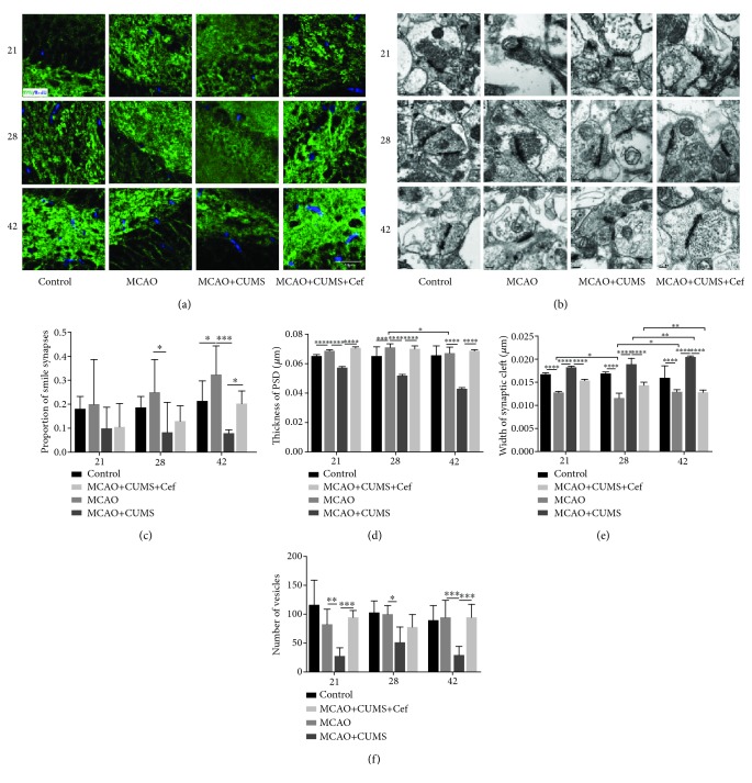
Figure 6.
Ceftriaxone improved synaptic plasticity in hippocampal DG of the CUMS-induced model rats. (a) Immunofluorescent images (1000×) of SYN (green) in the hippocampal DG on the 21st/28th/42nd day. Slices were also treated with BrdU to label new nuclei (blue). Scale bar: 40 μm. (b) Representative transmission electron images of synapses in DG (40000×) were pictured at the three time points. Scale bar: 0.2 μm. (c) Proportion of smile synapses of the CUMS-induced model rats was lower especially on the 42nd day. (d) Thickness of PSD in each synapse was smaller in the CUMS-induced model rats. (e) Average width of the synaptic cleft in synapses of the CUMS-induced model rats was significantly wider. (f) Decreased numbers of vesicles in synapses in the CUMS-induced model rats were reversed by ceftriaxone. ∗p < 0.05, ∗∗p < 0.005, ∗∗∗p < 0.0005, ∗∗∗∗p < 0.0001, two-way ANOVA (Tukey), n = 3.

