Figure 2.
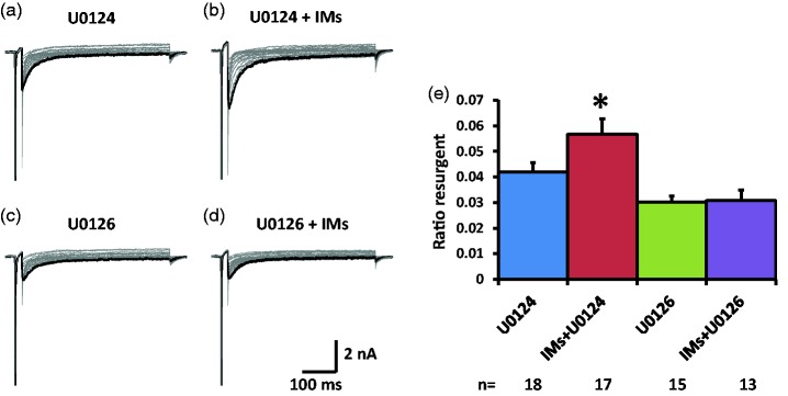
Effects of ERK inhibition on TTX-S resurgent currents in DRG neurons. Representative TTX-S, fast resurgent currents were recorded from medium DRG neurons ((a) to (d)). The resurgent currents were elicited by a series of repolarization voltage steps (+15 to −70 mV decreased by 5-mV steps, 450 ms) from a depolarization step (−100mV to +30 mV, 20 ms). ERK inhibitor U0126 (10 µM) and its inactive control U0124 (10 µM) were pretreated for 15 min in culture medium and were maintained in the recording chamber. (a and b) In the presence of U0124, the TTX-S resurgent currents were enhanced by a group of inflammatory mediators (IMs) including 1 µM bradykinin, 10 µM 5-HT, 10 µM histamine, 10 µM PGE2, and 5 µM ATP. (c and d) U0126 completely prevented the enhancing effects of inflammatory mediators on the TTX-S resurgent currents. Note that U0126 also significantly reduced TTX-S resurgent currents compared to U0124. (e) Statistical data of ratio resurgent currents were compared between groups with or without inflammatory mediators. Ratio resurgent currents were defined as peak resurgent currents normalized to peak TTX-S transient currents (maximal steady-state inactivation currents) in the same cells (the same below). The data were presented as mean ± standard error of the mean. Student’s t-test was used to compare the difference, *P < 0.05 (vs. U0124).

