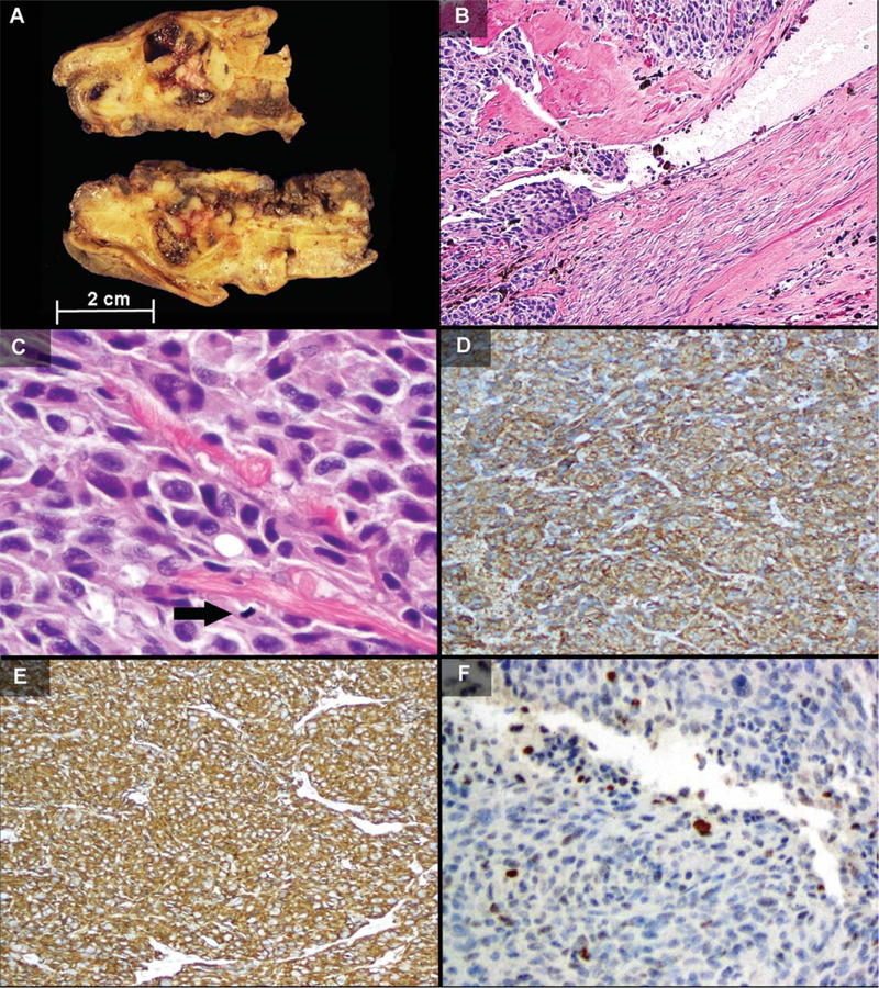
Figure 1.
Surgical pathology findings. A) Gross pathology of the recurrent post-imatinib gastric tumor showed multiple tan fleshy nodules with cystic hemorrhage within the gastric wall. B–F) The original resection demonstrated areas of lymphovascular invasion (B, H&E, 103), anaplasia with large tumor cells with multinucleation, intracytoplasmic vacuoles, and mitotic figure (arrow) (C, H&E, 40×), diffuse immunohistochemical positivity for KIT and CD34 (D and E, 10×), and patchy nuclear expression of MDM2 (F, 20×). [Color figure can be viewed in the online issue, which is available at wileyonlinelibrary.com.]

