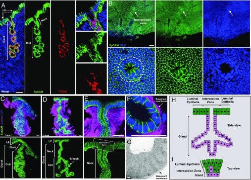
Fig. 1.
Characterization of mouse uterine endometrial epithelium. (A) Representative mouse uterine epithelia labeled with EpCAM antibody (epithelial marker, green), FOXA2 antibody (glandular-specific marker, red), and DAPI (nuclei, blue) in a wild-type uterine tissue section. Higher magnification of the glandular neck indicated in the dotted square in the Left merge panel is shown on the Right. (B, Upper) Intersection zone (indicated by arrow) between the luminal epithelial compartment and glandular tissue. (B, Lower) Higher magnification of the intersection zone circled on the Left. (C) A representative intact uterine epithelial unit dissected from adult wild-type uterus, stained with EpCAM antibody (green) and laminin antibody for basement membrane (magenta). (D) A representative loose uterine epithelial unit post manual dissociation of the coil compartment, stained as in C. (E) Higher magnification of glandular neck and intersection zone (circled by square) from the image in large dotted square in image D. (F) Higher magnification of glandular branch from the image in small dotted square in image D. (G) Microstructure of glandular tube by electron microscopy. Arrow indicates basement membrane. (H) Schematic of mouse uterine epithelial unit. (I) Cellular composition of intersection zone (Top view). Green indicates luminal cells, magenta indicates glandular cells. Data were collected from at least five adult wild-type mice for each independent experiment. (Scale bar, 2 μm in G and 50 μm in all other images.)

