Fig. 4.
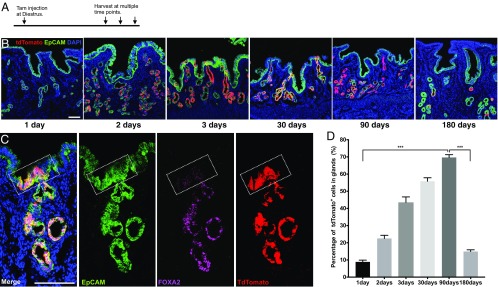
Founder cells of mixed clones do not exist in the glandular compartment. (A) Experimental design for tracing glandular cells. A single dosage of tamoxifen (4 mg per mouse) was given to adult Foxa2CreERT2;Rosa26Tom/+ mice at diestrus, then uteri were collected at estrus stage at multiple time points posttamoxifen injection for analysis. (B) Representative fluorescent images of uterine tissue after tracing of 1 d, 2 d, 3 d, 30 d, 90 d, or 180 d. Uterine tissues were collected from at least four mice at each time point for analysis. (C) Lineage marker tdTomato (red) is restricted to FOXA2+ cells (magenta). A representative tdTomato-labeled uterine epithelial unit stained with EpCAM (green) antibody, FOXA2 specifically labels glandular cells (magenta). The intersection zone is highlighted by a white square. (D) Percentage (%) of tdTomato+ glandular cells in total glandular cells at multiple time points posttracing. One-way ANOVA followed by Tukey’s test was applied here for the data assessment. ***P < 0.001. (Scale bar, 100 μm in all images.)

