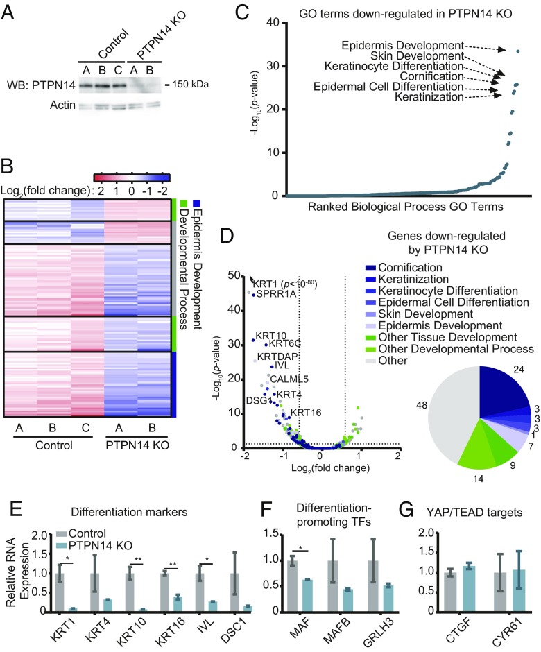
Fig. 5.
PTPN14 depletion impairs differentiation-related gene expression in primary human keratinocytes. Primary HFK were transduced with LentiCRISPRv2 lentiviral vectors encoding SpCas9 and nontargeting or PTPN14-directed sgRNAs and analyzed for changes in gene expression. (A) Cell lysates were subjected to SDS/PAGE/Western analysis and probed with anti-PTPN14 and antiactin antibodies. (B) PolyA selected RNA was analyzed by RNA-seq. Genes differentially expressed by ≥1.5-fold with P value ≤ 0.05 are displayed in the heat map. Color coding on the right side denotes whether genes are related to epidermis development (blue), other developmental processes (green), or neither (gray). (C) GO enrichment analysis of genes down-regulated in HFK-PTPN14 KO compared with HFK-control. (D) Volcano plot of gene-expression changes in HFK-control vs. HFK-PTPN14 KO. Dots colored by GO terms. Pie chart displays the fraction of genes down-regulated in the absence of PTPN14 that fall into enriched GO Terms. (E–G) Transcript abundance for selected genes in HFK-control and HFK-PTPN14 KO was measured by qRT-PCR detecting differentiation markers (E), differentiation promoting TFs (F), and YAP/TEAD targets (G). Bar graphs display the mean ± SD of two or three independent experiments. Statistical significance was determined by Welch’s t tests (*P < 0.05; **P < 0.01).

