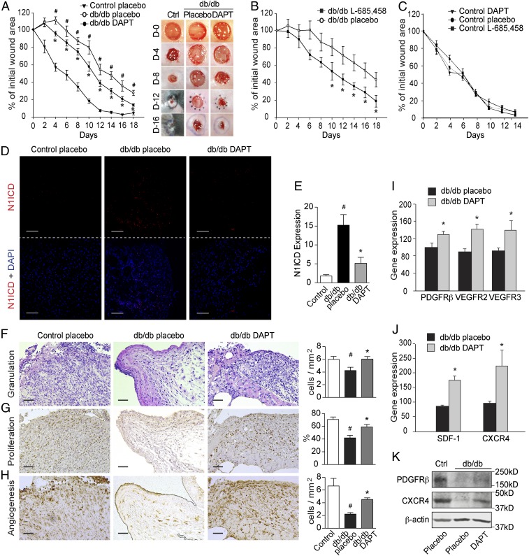
Fig. 4.
Inhibition of Notch signaling improves wound healing in diabetic mice. Full-thickness wounds were made on the dorsum of db/db and control mice, and the wounds were treated locally with DMSO (placebo) or GSIs DAPT (100 μM) or L-685,458 (100 μM) every second day. (A–C) The wound-healing rate was measured every second day and is shown as the percentage of the initial wound area (n = 5 per group). (A, Right) Representative wound images at days (D) 0, 4, 8, 12, and 16 during the healing process. (D) Representative images of immunohistochemistry staining for Notch1 ICD (N1ICD; red) and DAPI (blue) in wounds from control and db/db mice treated with placebo or DAPT. (E) Quantification results are shown (n = 9 or 10). (F–H) Levels of granulation, proliferation, and angiogenesis (microvessel density) were evaluated by histological analysis of hematoxylin and eosin staining (F), nuclear PCNA staining (G), and GSL-I isolectin B4 staining (H), respectively. Semiquantitative evaluations are presented in the histograms (n = 3). (I and J) The mRNA expression of PDGFRβ, VEGFR2, VEGFR3, SDF-1, and CXCR4 in diabetic wounds treated with DMSO (placebo) or DAPT (n = 10). (K) Western blotting results showing the expression of PDGFRβ and CXCR4 in control (Ctrl) and db/db mice treated with placebo or DAPT. (Scale bars: 50 µm.) #P < 0.05 (compared with control mice treated with placebo); *P < 0.05 (compared with db/db mice treated with placebo).

