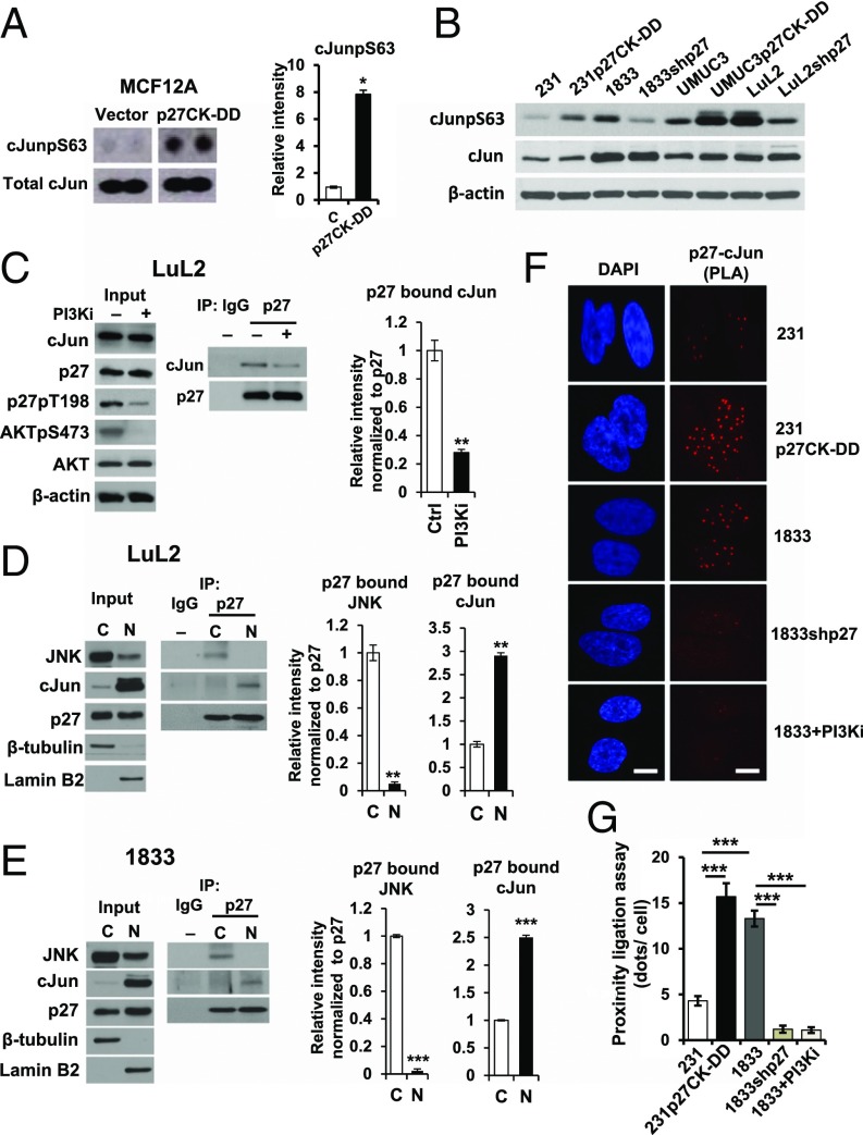
Fig. 3.
Association of cJun with p27 is increased by p27 phosphorylation. (A) Activated cJun (cJunpS63) in MCF12A controls (Vector) and MCF12Ap27CK−DD (p27CK−DD) by dot blot (Left) and densitometry values graphed as mean ± SEM (Right, *P < 0.05). (B) Western blots of cJun, cJunpS63, and β-actin in the indicated lines. (C) LuL2 treated for 48 h with the PI3K inhibitor PF1502 (PI3Ki). Western blots show input (Left) for p27 immunoprecipitation (IP) blotting to detect p27-associated cJun (Middle). Densitometric quantitation of p27-bound cJun (Right, **P < 0.01). See also SI Appendix, Fig. S2 A and B. (D and E) Nuclear (N) and cytoplasmic (C) fractions of LuL2 (D) and 1833 (E) were immunoblotted (Left), and p27-associated cJun and JNK were detected by IP blot (Middle) with densitometry (Right, **P < 0.01, ***P < 0.001). (F) In situ PLA shows p27/cJun complexes in the indicated lines and in 1833 treated with PF1502 (PI3Ki), indicated by red fluorescent dots. (G) Dots (mean ± SEM) graphed from triplicate PLAs; ANOVA with post hoc comparisons (***P < 0.001). (Scale bar: 10 µm.) See also EMSA data in SI Appendix, Fig. S2C.

